Refractive errors
Refractive errors of the eye include myopia, astigmatism, hypermetropia and presbyopia.
Glaucoma
Glaucoma is a degenerative neuropathy that destroys the nerve fibers of the optic nerve.
Refractive errors of the eye include myopia, astigmatism, hypermetropia and presbyopia.
Glaucoma is a degenerative neuropathy that destroys the nerve fibers of the optic nerve.
Refractive errors of the eye include myopia, astigmatism, hypermetropia and presbyopia.
The characteristic of refractive errors is that the rays of light do not all end up together in the center of vision at the back of the eye as is normal, due to structural abnormalities in the optical media. Depending on the error, the rays may end up in front of the focal point that should be (myopia), behind the focal point that should be (hyperopia) or in 2 lines perpendicular to each other in front or behind the focal point that should be (astigmatism).
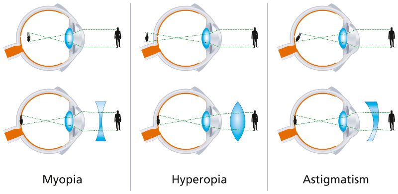
In presbyopia, the crystalline lens of the eye has difficulty zooming in when the person looks closely, i.e. it curves, due to hardening of its material after the age of 40, so the person cannot focus clearly on near objects.
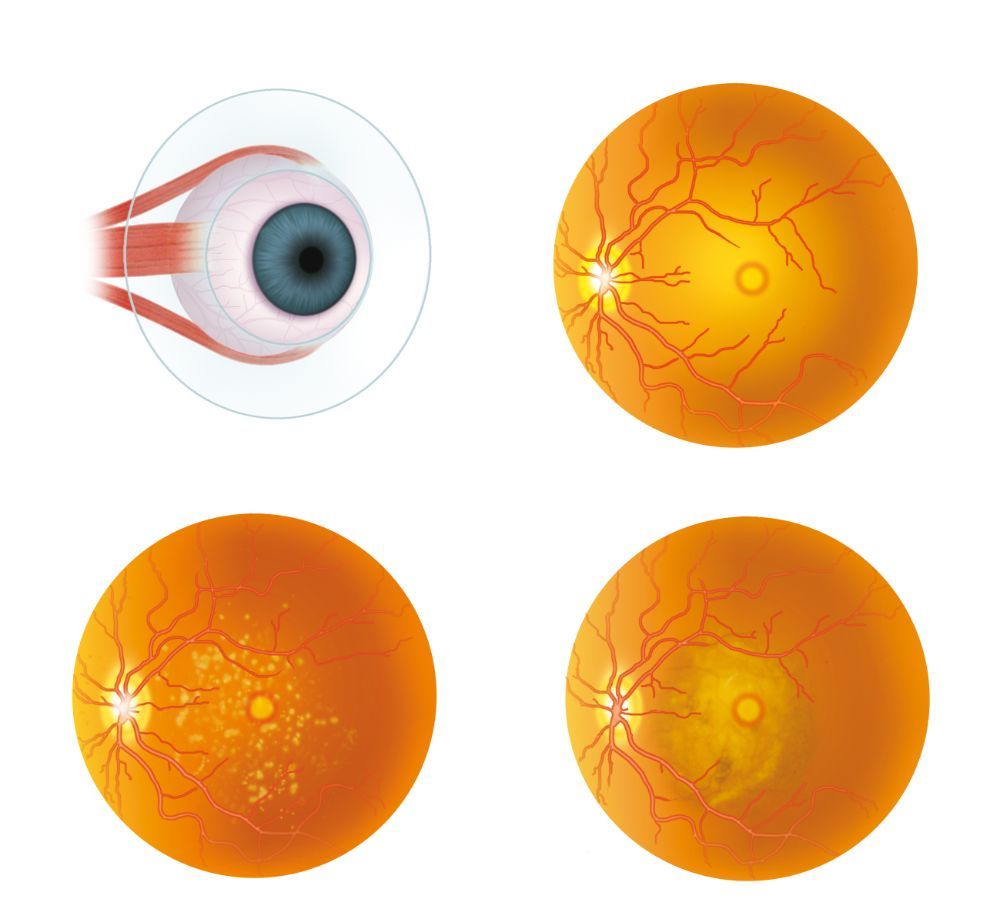
The retina is a structure located at the back of the eye internally.
The main diseases that affect the retina are:
The office is equipped with a latest technology OCT machine and digital angiography, where contrast injection is not required from the vein as was done with the old conventional fluoro angiography.
Glaucoma is a degenerative neuropathy that destroys the nerve fibers of the optic nerve, resulting in damage to the peripheral vision in the early stages, and in later more advanced stages, damage to the central vision as well as blindness.
The major risk factor for glaucoma and the only one that can be controlled by drugs is intraocular pressure.
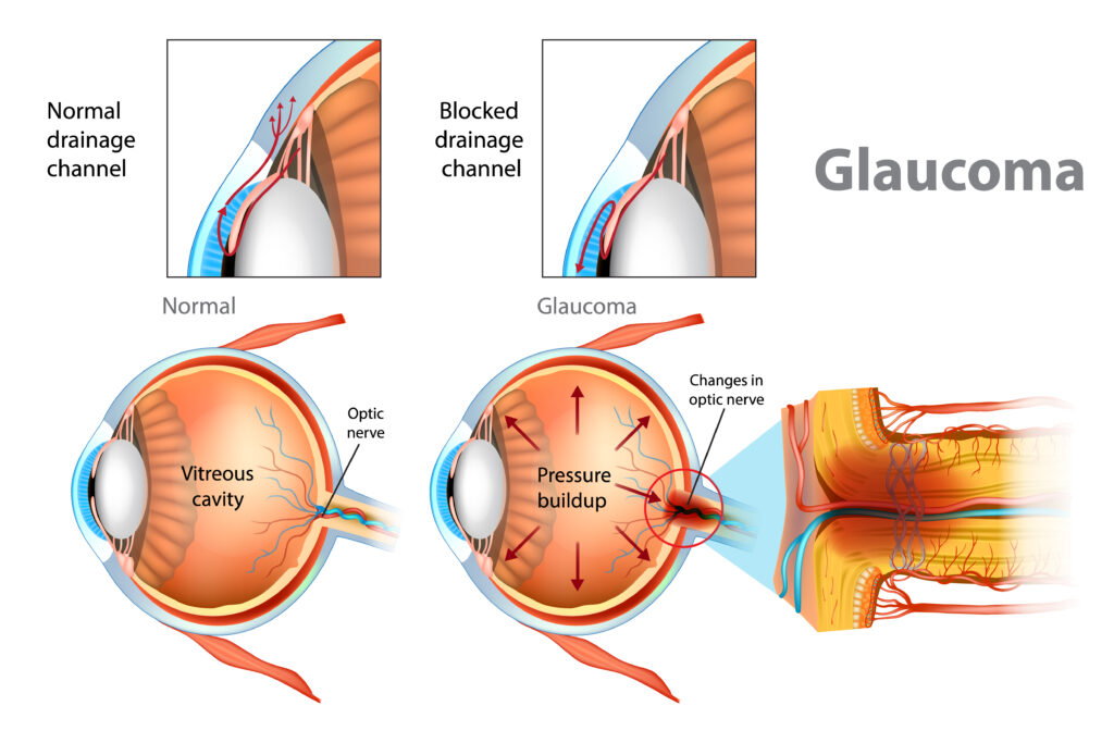
During fundoscopy we see the of the optic nerve head, and the large damages in it, if any, are visible, but smaller damages are not apparent.
In order to check in more detail whether there is loss of nerve fibers and damage to peripheral vision, the visual field machine (detection of functional vision loss) and tomography of the optic nerve (nerve OCT) are used to detect the anatomical loss of nerve fibers.
